|
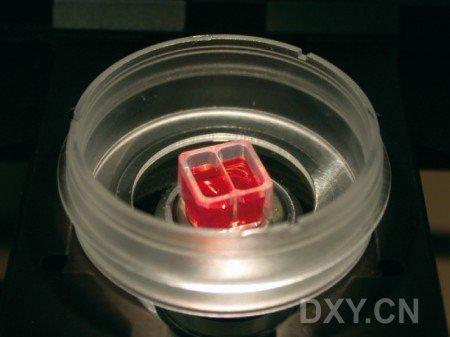
ibidi Culture Insert 系列

ibidi公司根据伤口愈合和细胞迁移实验的要求设计,它较之传统的划痕实验,有以下优点,
首先:可以确定划痕的宽度和面积,因此实验可重复性好;
其次:可移除的插入元件(Culture-Insert)对玻片表面包被的几乎没有损伤,因此不影响后期gap处细胞的生长和迁移;
最后:配合ibidi加热和孵育系统,可以进行清晰,长期的活细胞显微拍摄;
使用方法
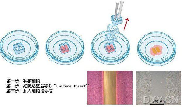
1. Sample Preparation——样品准备

当放入细胞培养表面,ibidi-Culture-Insert提供两个中间有500um的间隙的独立培养室。细胞在两个培养室生长,之后将这个硅胶元件从表面移除。移除后,由于中间的隔断壁,在培养表面形成规则的细胞补丁,两块补丁中间有非常规则,宽度一致的缝隙。
由于这个独特的设计,ibidi-Culture-Inser自身粘附在培养表面,这样的设计完全避免了细胞在隔断壁下生长。从表面移除后,这个细胞间隙(也就是伤口)会没有任何污染和改变,且培养表面的包被也不会被破坏。通过这个方法,一个没有任何细胞覆盖且非常规则的区域。
2. Microscopy/显微观察

3. Image Analysis / 显微分析
4 . Quantitative and Statistical Analysis/定量和统计分析
Data Acquisition/数据采集:
显微镜用于评估伤口愈合的过程。根据你的关注点和兴趣,不仅可以视频显微镜,还可以进行定点的成像观察。ibidi 加热与孵育系统可以直接在培养的条件下进行活细胞成像,不用将样品反复的从孵育室移至显微镜上。
Data Analysis/数据分析:
ibidi的伤口愈合成像分析解决方案可以评估2D细胞迁移,通过4步就可以得到结果。

网页自动分析软件--WimScratch软件可以将您手机的数据在几分钟内自动生成结果,你只需上传数据到图像分析平台,结果将通过电子邮件即时发送给您。
分析数据包括:
a) 细胞覆盖面积
b) 终止速度(平均≥5个图像)
c) 加速特征(平均≥5个图像)
d) 概览图
e) 中间接合处的近似值(平均≥5个图像)
|
规格/Specifications:
|
|
Number of wells
|
2
|
|
Outer dimensions (w x l x h) in mm
|
8.4 x 8.4 x 5
|
|
Volume per well
|
70 ul
|
|
Growth area per well
|
0.22 cm2
|
|
Width of cell free gap
|
500 um +/- 50 um
|
|
Material
|
Biocompatible silicone
|
|
Bottom
|
No bottom - sticky underside
|
货号:
|
Cat. No
|
Description
|
Pcs./Box
|
|
Eubio80206
|
u-Dish 35 mm, low Culture-Insert, ready to use in a u-Dish 35 mm, low wall, ibiTreat, tissue culture treated, sterile
|
30
|
|
Eubio80201
|
u-Dish 35 mm, low Culture-Insert, ready to use in a u-Dish 35 mm, low wall, hydrophobic, uncoated, sterile
|
30
|
|
Eubio 81176
|
u-Dish 35 mm, high Culture-Insert, Culture-Inserts ready to use in a u-Dish 35 mm, high wall, ibiTreat, tissue culture treated, sterile
|
30
|
|
Eubio 81171
|
u-Dish 35 mm, high Culture-Insert, Culture-Inserts ready to use in a u-Dish 35 mm, high wall, hydrophobic, uncoated, sterile
|
30
|
|
Eubio 80241
|
Culture-Insert 24, a 24 well plate with 24 ready to use Culture-Inserts, tissue culture treated, polystyrene, sterile
|
3
|
|
Eubio 80209
|
25 Culture-Inserts for self-insertion, sterile, in a 10 cm transport dish
|
25
|
|